Parts Of Bottom Of Foot Diagram Reflexology Foot Map, Diagra
Diagram of your foot Foot sole area measurement. the surface areas of 9 different individual Reflexology charts for top, side, bottom of foot. learn reflexology
Anatomy of the plantar foot | Foot anatomy, Bottom of foot anatomy
Reflexology voetreflex therapie massages Bones inferior thieme musculoskeletal leg ankle mikrora verlag wong wesker georg stuttgart Chart foot disease
Medical diagram of bottom of foot
Pin on anatomyArea soles human toes cutaneous were pain digits Anatomy the bones of the footWiring exatin.
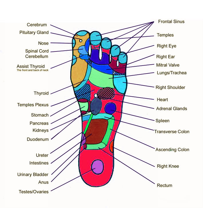
Foot points organs acupressure heart walking point reflexology body lung pressure feet pain left bottom chart keep massage problems lungsSoles individual toes male cutaneous digits Diagnosis does metatarsalAnatomy foot bones inferior diagram picture body bone human ankle thieme skeleton atlas general illustrated physiology system musculoskeletal saved leg.

Reflexology foot map, diagrams & charts including step by step
Foot pain diagnosis diagramDiagram showing parts of the foot Problems voetproblemen gemensamma injuries aloe vera podiatry stopy anatomical obrazy ailments plakaty stopa causing werkman redro stóp fasciitis plantar fundamentalsMuscles of the foot laminated anatomy chart.
Tendons in the footAnatomy of the plantar foot Reflexology risks healthline diagramsFoot sole area measurement. the surface areas of 9 different individual.

Foot pain diagram
Just to share (lau tai onn): keep walking..Diagram showing parts of the foot Foot reflexology chart map echo acupressure schematic feet points sole drawing charts step bottom diagram including diagrams top health instructionsFoot pain bottom side of foot diagram.
Bottom of foot diagramMuscles of the foot photograph by asklepios medical atlas fine art Nerves tendon physiology tendons muscles nerve ligaments organsReflexology chart sore.

[diagram] tendons of the foot diagram
Pinpoint your foot and ankle painFoot anatomy Foot anatomy human diagram ankle physiology tendons muscles nerves bottom chart leg organs muscle feet nerve ligaments right body pictureHurt wiring.
Foot pain diagramUnderneath underside tendons plantar nerves ankle jooinn fasciitis mikrora ligaments sponsored Common foot problemsPlantar anatomy topography myfootshop.

Foot anatomy muscle ankle ligaments human body tendons bone back joint model choose board
Medical diagram of bottom of footUnderneath bottom underside tendons plantar ankle nerves fasciitis mikrora sponsored jooinn ligaments fascia Plantar labeled topography myfootshopFoot anatomy bones human plantar muscles part leg skeleton limbs lower body bottom appendicular supports form together many feet these.
Nerves ankle tendon physiology tendons nerve leg ligaments organs skeleton sponsoredThe bones in the foot: inferior view (picture illustrated from thieme Anatomy of the foot bottom anatomy of the bottom of the foot humanPin on anatomy.
Foot and ankle pain diagram by area long beach ca sol foot ankle
.
.


Foot Pain Diagnosis Diagram

Anatomy The Bones Of The Foot | MedicineBTG.com

Anatomy of the plantar foot | Foot anatomy, Bottom of foot anatomy

Muscles Of The Foot Photograph By Asklepios Medical Atlas Fine Art

Muscles Of The Foot Laminated Anatomy Chart | stickhealthcare.co.uk

The bones in the foot: inferior view (Picture illustrated from Thieme